
Question 1
- What is a microscope?
- What does ‘microscopic’ mean?
- What is microscopy?
Question 2
State the three components of the cell theory.
Question 3
Distinguish between the following types of microscopes.
- A light microscope and an electron microscope.
- A compound microscope and a stereo microscope.
- A monocular microscope and a binocular microscope.
- A scanning electron microscope and a transmission electron microscope.
Question 4
Match the components A-J on the following diagram of a compound microscope with the correct labels from the word list below.
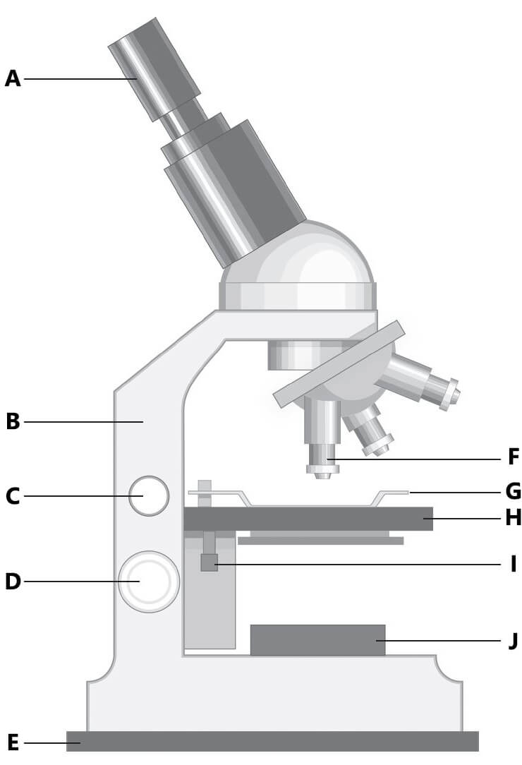
arm • base • coarse focus knob • eyepiece • fine focus knob • lamp • objective lens • stage • stage clips • stage controls
Question 5
Describe the function of the following parts of a compound microscope.
- Eyepiece (ocular lens)
- Objective lens
- Coarse focus knob
- Fine focus knob
- Stage
- Stage clips
- Stage controls
- Lamp
Question 6
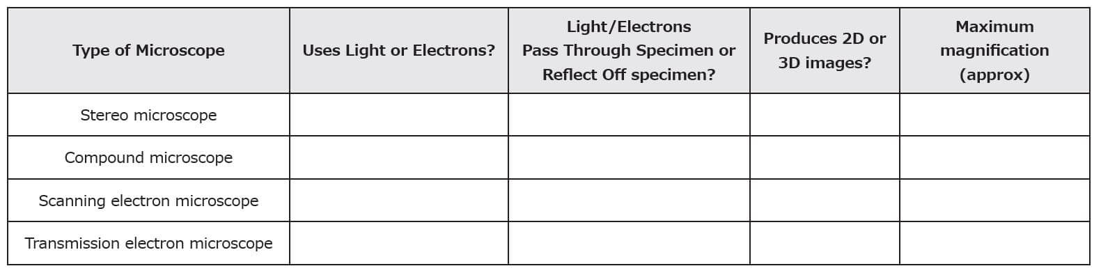
Complete the following table.
Question 7
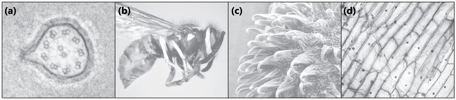
State the type of microscope used to produce the following images. (Select from the four types listed in Q6.)
Question 8
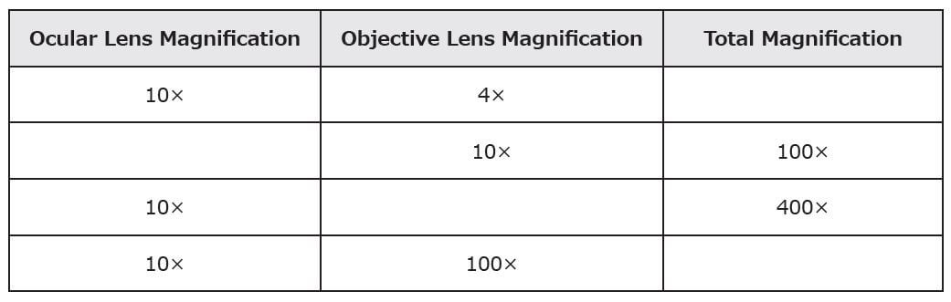
Complete the following table of magnification.
Question 9
The image below shows red blood cells viewed under a microscope.
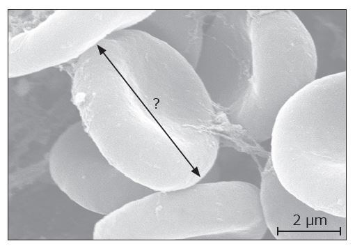
- What length does the scale bar represent? (This is known as the ‘actual size’.)
- Measure the length of the scale bar (to the nearest mm) using your ruler. (This is known as the ‘image size’.)
- Convert your answer for (b) into μm. (1 mm = 1000 μm.)
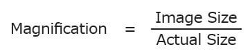
- Calculate the magnification of the image using the formula below:
- Calculate the actual diameter of a red blood cell based on the arrow labelled with a question mark.